Mixtures are heterogeneous forms of matter. Mixtures are composed of variable proportions of molecules and atoms.
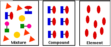
The composition of a mixture is variable with each components retaining its characteristic properties. Its components are easily separated. Examples of Mixtures: soil, ocean water and other solutions, air, the cytosol of a cell
n contrast, compounds are homogeneous forms of matter. Their constituent elements (atoms and/or ions) are always present in fixed proportions . Properties of compounds include
- The relative proportions of the elements in a compound are fixed.
- The components of a compound do not retain their individual properties. Both sodium and chlorine are poisonous; their compound, table salt (NaCl) is absolutely essential to life.
- It takes large inputs of energy to separate the components of a compound.
Examples of Compounds
- water (H2O)
- table salt (NaCl)
- sucrose (table sugar, C12H22O11)
Separating the Components of a Mixture
Most laboratory work in biology requires the use of techniques to separate the components of mixtures. This is done by exploiting some property that distinguishes the components, such as their relative
- size
- density
- solubility
- electrical charge
Dialysis
Dialysis is the separation of small solute molecules or ions (e.g., glucose, Na+, Cl-) from macromolecules (e.g., starch) by virtue of their differing rates of diffusion through a differentially permeable membrane.
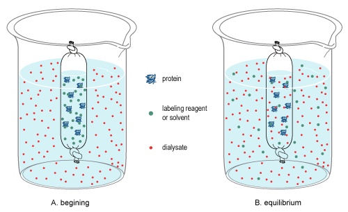
As shown in Figure \(\PageIndex{1}\), the cellophane used to construct a bag is perforated with tiny pores that permit ions and small molecules to pass through, but exclude molecules with molecular weights greater than about 12,000. If a cellophane bag is mixed with a mixture of sugar and starch and place it in salt water, the sugar molecules (teal dots) will diffuse out into the water until equilibrium is reached; i.e., until their concentrations are equal on both sides of the membrane. Similarly, the salt (red dots) will diffuse into the bag. However, because of their large size, all the starch (big blue disks) will be retained within the tubing.
Chromatography
Chromatography is the term used for several techniques for separating the components of a mixture. The different types of chromatography techniques used are: paper chromatography, exclusion chromatography, and affinity chromatography.
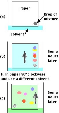
Paper chromatography technique provides an easy way to separate the components of a mixture. A drop of mixture is placed in one corner of a square of absorbent paper.
- One edge of the paper is immersed in a solvent. (a)
- The solvent migrates up the sheet by capillary attraction.
- As it does so, the substances in the drop are carried along at different rates. (b)
- Each compound migrates at a rate that reflects
- the size of its molecule and
- its solubility in the solvent.
- After a second run at right angles to the first (often using a different solvent), the various substances will be spread out at distinct spots across the sheet, forming a chromatogram. (c)
- The identity of each spot can be determined by comparing its position with the position occupied by known substances under the same conditions.
- In many cases, a fragment of the paper can be cut away from the sheet and chemical analysis run on the tiny amount of substance in it.
Autoradiography
If the mixture contains molecules that have been labeled with a radioactive isotope, these can be located by placing the chromatogram next to a sheet of X-ray film. The location of dark spots on the developed film (because of radiation emitted by the isotope) can be correlated with the position of the substances on the chromatogram.
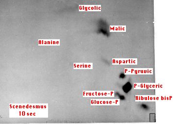
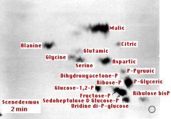
The above figures (courtesy of Dr. James A. Bassham) show autoradiograms of the type that were essential in working out the dark reactions of photosynthesis. The dark spots show the radioactive compounds produced after 10 secs (left) and 2 minutes (right) of photosynthesis by the green alga Scenedesmus. The alga was supplied with carbon dioxide labeled with 14C, a radioactive isotope of carbon.
- At 10 seconds, most of the radioactivity is found in 3-phosphoglyceric acid ("P-Glyceric").
- At 2 minutes, phosphorylated 6-carbon sugars (glucose and fructose) have been synthesized as well as a number of amino acids.
The small rectangle and circle (lower right-hand corners) mark the spots where the cell extract was applied.
Exclusion chromatography
One of the most common problems in biochemical research is to separate the many components — usually macromolecules — in cell extracts and the like. Methods for separating the components of a mixture exploit such differences as size, electrical charge, and solubility in different solvents. of the molecules in it. One example: Electrophoresis which separates such macromolecules as proteins and DNA by their charge (and sometimes size as well).
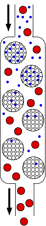
Exclusion chromatography separates molecules on the basis of size. A column is filled with semi-solid beads of a polymeric gel that will admit ions and small molecules (blue) into their interior but not large ones (shown in red). When a mixture of molecules and ions dissolved in a solvent is applied to the top of the column, the smaller molecules (and ions) are distributed through a larger volume of solvent than is available to the large molecules. Consequently, the large molecules move more rapidly through the column, and in this way the mixture can be separated (fractionated) into its components. The porosity of the gel can be adjusted to exclude all molecules above a certain size. Sephadex and sepharose are trade names for gels that are available commercially in a broad range of porosities.
Affinity chromatography
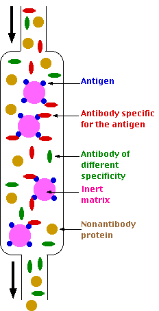 The goal of affinity chromatography is to separate all the molecules of a particular specificity from the whole gamut of molecules in a mixture such as a blood serum. For example, the antibodies in a serum sample specific for a particular antigenic determinant can be isolated by the use of affinity chromatography.
The following steps are performed to achieve that:
Step 1
An immunoadsorbent is prepared. This consists of a solid matrix to which the antigen (shown in blue) has been coupled (usually covalently). Agarose, sephadex, derivatives of cellulose, or other polymers can be used as the matrix.
Step 2
The serum is passed over the immunoadsorbent. As long as the capacity of the column is not exceeded, those antibodies in the mixture specific for the antigen (shown in red) will bind (noncovalently) and be retained. Antibodies of other specificities (green) and other serum proteins (yellow) will pass through unimpeded.
Step 3
Elution. A reagent is passed into the column to release the antibodies from the immunoadsorbent. Buffers containing a high concentration of salts and/or low pH are often used to disrupt the noncovalent interactions between antibodies and antigen. A denaturing agent, such as 8 M urea, will also break the interaction by altering the configuration of the antigen-binding site of the antibody molecule.
Another, gentler, approach is to elute with a soluble form of the antigen. These compete with the immunoadsorbent for the antigen-binding sites of the antibodies and release the antibodies to the fluid phase.
Step 4
Dialysis. The eluate is then dialyzed against, for example, buffered saline in order to remove the reagent used for elution.
Electrophoresis
Electrophoresis uses a direct electric current to separate the components of a mixture by the differing electrical charge.
Example of Electrophoresis
Proteins in blood serum can be separated by electrophoresis.
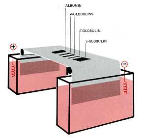
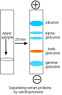
- A drop of serum is applied in a band to a thin sheet of supporting material, like paper, that has been soaked in a slightly-alkaline salt solution.
- At pH 8.6, which is commonly used, all the proteins are negatively charged, but some more strongly than others.
- A direct current can flow through the paper because of the conductivity of the buffer with which it is moistened.
- As the current flows, the serum proteins move toward the positive electrode.
- The stronger the negative charge on a protein, the faster it migrates.
- After a time (typically 20 min), the current is turned off and the proteins stained to make them visible (most are otherwise colorless).
- The separated proteins appear as distinct bands.
- The most prominent of these and the one that moves closest to the positive electrode is serum albumin.
- The other proteins are the various serum globulins.
Pure Substances
Some of the pure substances isolated from mixtures cannot be further broken down. Oxygen (O2) is an example. It is one of the elements; the fundamental building blocks of matter. Most pure substances are compounds. Table salt, sodium chloride (NaCl), is an example; water (H2O) is another. If we pass an electrical current through molten NaCl, two new substances will be formed:
- sodium, a shiny metal so reactive that it must be stored out of contact with the air
- chlorine, a yellowish poisonous gas.
In this operation, a compound has been decomposed into its constitutive elements. Note the differences between separating the components of a mixture and those of a compound. The decomposition of NaCl required a large input of energy since the strong ionic bonds holding the Na and Cl atoms together must be broken. The ratio of the weights of the two products are always 23 parts of sodium to 35.5 parts of chlorine. This reflects the invariance of the ratio (1:1 in this case) of the number of atoms in a compound and the relative weights (23:35.5) of the atoms in table salt. The properties of the components of the compound are not the same as those of the compound itself. Both sodium and chlorine are hazardous to life; their compound, sodium chloride, is a vital ingredient of all animal diets.