Apoptosis is a process of programmed cell death that occurs in multicellular organisms. There are two ways in which cells die: (1) They are killed by injurious agents or (2) they are induced to commit suicide.
Death by injury
Cells that are damaged by injury, such as by mechanical damage or exposure to toxic chemicals undergo a characteristic series of changes. They (and their organelles like mitochondria) swell (because the ability of the plasma membrane to control the passage of ions and water is disrupted). The cell contents leak out, leading to inflammation of surrounding tissues.
Death by Suicide
Cells that are induced to commit suicide:
- shrink
- develop bubble-like blebs on their surface
- have the chromatin (DNA and protein) in their nucleus degraded
- have their mitochondria break down with the release of cytochrome c
- break into small, membrane-wrapped, fragments
- release (at least in mammalian cells) ATP and UTP
- These nucleotides bind to receptors on wandering phagocytic cells like macrophages and dendritic cells and attract them to the dying cells (a "find-me" signal")
- The phospholipid phosphatidylserine, which is normally hidden in the inner layer of the plasma membrane, is exposed on the surface
- This "eat me" signal is bound by other receptors on the phagocytes which then engulf the cell fragments
- The phagocytic cells secrete cytokines that inhibit inflammation (e.g., IL-10 and TGF-β)
The pattern of events in death by suicide is so orderly that the process is often called programmed cell death or PCD. The cellular machinery of programmed cell death turns out to be as intrinsic to the cell as, say, mitosis. Programmed cell death is also called apoptosis. (There is no consensus yet on how to pronounce it; some say APE oh TOE sis; some say uh POP tuh sis.)
Why should a cell commit suicide?
There are two different reasons.
1. Programmed cell death is as needed for proper development as mitosis is.
Examples:
- The resorption of the tadpole tail at the time of its metamorphosis into a frog occurs by apoptosis.
- The formation of the fingers and toes of the fetus requires the removal, by apoptosis, of the tissue between them.
- The sloughing off of the inner lining of the uterus (the endometrium) at the start of menstruation occurs by apoptosis.
- The formation of the proper connections (synapses) between neurons in the brain requires that surplus cells be eliminated by apoptosis.
- The elimination of T cells that might otherwise mount an autoimmune attack on the body occurs by apoptosis.
- During the pupal stage of insects that undergo complete metamorphosis, most of the cells of the larva die by apoptosis thus providing the nutrients for the development of the structures of the adult.
2. Programmed cell death is needed to destroy cells that represent a threat to the integrity of the organism.
Examples:
- Cells infected with viruses
- One of the methods by which cytotoxic T lymphocytes (CTLs) kill virus-infected cells is by inducing apoptosis and some viruses mount countermeasures to thwart it.
- Cells of the immune system
- As cell-mediated immune responses wane, the effector cells must be removed to prevent them from attacking body constituents. CTLs induce apoptosis in each other and even in themselves. Defects in the apoptotic machinery is associated with autoimmune diseases such as systemic lupus erythematosus and rheumatoid arthritis.
- Cells with DNA damage
- Damage to its genome can cause a cell
- to disrupt proper embryonic development leading to birth defects
- to become cancerous.
- Cancer cells
- Radiation and chemicals used in cancer therapy induce apoptosis in some types of cancer cells.
What makes a cell decide to commit suicide?
The balance between the withdrawal of positive signals; that is, signals needed for continued survival, and the receipt of negative signals.
Withdrawal of positive signals
The continued survival of most cells requires that they receive continuous stimulation from other cells and, for many, continued adhesion to the surface on which they are growing. Some examples of positive signals: growth factors for neurons and Interleukin-2 (IL-2), an essential factor for the mitosis of lymphocytes
Receipt of negative signals
- increased levels of oxidants within the cell
- damage to DNA by these oxidants or other agents like ultraviolet light, X-rays and chemotherapeutic drugs
- accumulation of proteins that failed to fold properly into their proper tertiary structure
- molecules that bind to specific receptors on the cell surface and signal the cell to begin the apoptosis program. These death activators include:
- Tumor necrosis factor-alpha (TNF-α) that binds to the TNF receptor
- Lymphotoxin (also known as TNF-β) that also binds to the TNF receptor
- Fas ligand (FasL), a molecule that binds to a cell-surface receptor named Fas (also called CD95)
The Mechanisms of Apoptosis
There are 3 different mechanisms by which a cell commits suicide by apoptosis.
- Generated by signals arising within the cell
- Triggered by death activators binding to receptors at the cell surface:
- TNF-α
- Lymphotoxin
- Fas ligand (FasL)
- Triggered by dangerous reactive oxygen species
Apoptosis triggered by internal signals
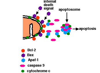
- In a healthy cell, the outer membranes of its mitochondria display the protein Bcl-2 on their surface. Bcl-2 inhibits apoptosis.
- Internal damage to the cell
- causes a related protein, Bax, to migrate to the surface of the mitochondrion where it inhibits the protective effect of Bcl-2 and inserts itself into the outer mitochondrial membrane punching holes in it and causing
- cytochrome c to leak out.
- The released cytochrome c binds to the protein Apaf-1 ("apoptotic protease activating factor-1").
- Using the energy provided by ATP, these complexes aggregate to form apoptosomes. The apoptosomes bind to and activate caspase-9. Caspase-9 is one of a family of over a dozen caspases. They are all proteases. They get their name because they cleave proteins — mostly each other — at aspartic acid (Asp) residues.
- Caspase-9 cleaves and, in so doing, activates other caspases (caspase-3 and -7).
- The activation of these "executioner" caspases creates an expanding cascade of proteolytic activity (rather like that in blood clotting and complement activation) which leads to
- digestion of structural proteins in the cytoplasm,
- degradation of chromosomal DNA
- phagocytosis of the cell
Apoptosis triggered by external signals
- Fas and the TNF receptor are integral membrane proteins with their receptor domains exposed at the surface of the cell
- Binding of the complementary death activator (FasL and TNF respectively) transmits a signal to the cytoplasm that leads to the activation of caspase 8
- Caspase 8 (like caspase 9) initiates a cascade of caspase activation leading to phagocytosis of the cell.
- Example: When cytotoxic T cells recognize (bind to) their target,
- They produce more FasL at their surface.
- This binds with the Fas on the surface of the target cell leading to its death by apoptosis.
The early steps in apoptosis are reversible — at least in C. elegans. In some cases, final destruction of the cell is guaranteed only with its engulfment by a phagocyte.
Apoptosis-Inducing Factor (AIF)
Neurons, and perhaps other cells, have another way to self-destruct that — unlike the two paths described above — does not use caspases. Apoptosis-inducing factor (AIF) is a protein that is normally located in the intermembrane space of mitochondria. When the cell receives a signal telling it that it is time to die, AIF is released from the mitochondria (like the release of cytochrome c in the first pathway). It migrates into the nucleus and binds to DNA, which triggers the destruction of the DNA and cell death.
Apoptosis and Cancer
Some viruses associated with cancers use tricks to prevent apoptosis of the cells they have transformed.
- Several human papilloma viruses (HPV) have been implicated in causing cervical cancer. One of them produces a protein (E6) that binds and inactivates the apoptosis promoter p53.
- Epstein-Barr Virus (EBV), the cause of mononucleosis and associated with some lymphomas
- produces a protein similar to Bcl-2
- produces another protein that causes the cell to increase its own production of Bcl-2. Both these actions make the cell more resistant to apoptosis (thus enabling a cancer cell to continue to proliferate).
Even cancer cells produced without the participation of viruses may have tricks to avoid apoptosis.
- Some B-cell leukemias and lymphomas express high levels of Bcl-2, thus blocking apoptotic signals they may receive. The high levels result from a translocation of the BCL-2 gene into an enhancer region for antibody production.
- Melanoma (the most dangerous type of skin cancer) cells avoid apoptosis by inhibiting the expression of the gene encoding Apaf-1.
- Some cancer cells, especially lung and colon cancer cells, secrete elevated levels of a soluble "decoy" molecule that binds to FasL, plugging it up so it cannot bind Fas. Thus, cytotoxic T cells (CTL) cannot kill the cancer cells by the mechanism shown above.
- Other cancer cells express high levels of FasL, and can kill any cytotoxic T cells (CTL) that try to kill them because CTL also express Fas (but are protected from their own FasL).
Apoptosis in the Immune System
The immune response to a foreign invader involves the proliferation of lymphocytes — T and/or B cells. When their job is done, they must be removed leaving only a small population of memory cells. This is done by apoptosis. Very rarely humans are encountered with genetic defects in apoptosis. The most common one is a mutation in the gene for Fas, but mutations in the gene for FasL or even one of the caspases are occasionally seen. In all cases, the genetic problem produces autoimmune lymphoproliferative syndrome or ALPS.
Features
- an accumulation of lymphocytes in the lymph nodes and spleen greatly enlarging them.
- the appearance of clones that are autoreactive; that is, attack "self" components producing such autoimmune disorders as
- hemolytic anemia
- thrombocytopenia
- the appearance of lymphoma — a cancerous clone of lymphocytes.
In most patients with ALPS, the mutation is present in the germline; that is, every cell in their body carries it. In a few cases, however, the mutation is somatic; that is, has occurred in a precursor cell in the bone marrow. These later patients are genetic mosaics — with some lymphocytes that undergo apoptosis normally and others that do not. The latter tend to out-compete the former and grow to become the major population in the lymph nodes and blood.
Apoptosis and Organ Transplants
For many years it has been known that certain parts of the body such as the anterior chamber of the eye and the testes are "immunologically privileged sites". Antigens within these sites fail to elicit an immune response. It turns out that cells in these sites differ from the other cells of the body in that they express high levels of FasL at all times. Thus antigen-reactive T cells, which express Fas, would be killed when they enter these sites. (This is the reverse of the mechanism described above.)
This finding raises the possibility of a new way of preventing graft rejection. If at least some of the cells on a transplanted kidney, liver, heart, etc. could be made to express high levels of FasL, that might protect the graft from attack by the T cells of the host's cell-mediated immune system. If so, then the present need for treatment with immunosuppressive drugs for the rest of the transplant recipient's life would be reduced or eliminated. So far, the results in animal experiments have been mixed. Allografts engineered to express FasL have shown increased survival for kidneys, but not for hearts or islets of Langerhans.
Apoptosis in Plants
Plants, too, can turn on a system of programmed cell death; for example, in an attempt to halt the spread of virus infection. The mechanism differs from that in animals although it, too, involves a protease that — like caspases — cleaves other proteins at Asp (and Asn) residues. Activation of this enzyme destroys the central vacuole, which is followed by disintegration of the rest of the cell.