Collagens are insoluble, extracellular glycoproteins found in all animals. They are the most abundant proteins in the human body and are essential structural components of all connective tissues such as cartilage, bone, tendons, ligaments, fascia, skin. Gelatin is solubilized collagen. 29 types of collagens have been found in humans and the major ones are:
- Type I. The chief component of tendons, ligaments, and bones.
- Type II. Represents more than 50% of the protein in cartilage and is the major component of the vitreous body of the eye. It is also used to build the notochord of vertebrate embryos.
- Type III. Strengthens the walls of hollow structures like arteries, the intestine, and the uterus.
- Type IV. Forms the basal lamina of epithelia. (The basal lamina is often called the basement membrane, but is not related to lipid bilayer membranes.) A meshwork of Type IV collagens provides the filter for the blood capillaries and the glomeruli of the kidneys.
The other 25 types are probably equally important, but they are much less abundant.
Collagens Structure
The basic unit of collagens is a polypeptide consisting of the repeating sequence
n
where X is often proline (Pro) and Y is often hydroxyproline (proline to which an -OH group is added after synthesis of the polypeptide). The resulting molecule twists into an elongated, left-handed helix (NOT an alpha helix).
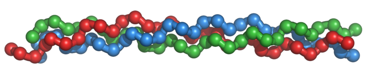
A single collagen molecule, tropocollagen, is used to make up larger collagen aggregates, such as fibrils. It is approximately 300 nm long and 1.5 nm in diameter, and it is made up of three polypeptide strands (called alpha peptides, see step 2), each of which has the conformation of a left-handed helix (Figure 3.21.1). These three left-handed helices are twisted together into a right-handed triple helix or "super helix", a cooperative quaternary structure stabilized by many hydrogen bonds.
When synthesized, the N- terminal and C- terminal of the polypeptide have globular domains, which keep the molecule soluble. As they pass through the endoplasmic reticulum (ER) and Golgi apparatus,
- The molecules are glycosylated.
- Hydroxyl (-OH) groups are added to the "Y" amino acid.
- S-S bonds link three chains covalently.
- The three molecules twist together to form a triple helix.
In some collagens (e.g., Type II), the three molecules are identical (the product of a single gene). In other collagens (e.g., Type I), two polypeptides of one kind (gene product) assemble with a second, quite similar, polypeptide, that is the product of a second gene.
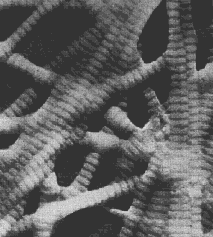
When the triple helix is secreted from the cell (usually by a fibroblast), the globular ends are cleaved off. The resulting linear, insoluble molecules assemble into collagen fibers. They assemble in a staggered pattern that gives rise to the striations seen in the above electron micrograph. Type IV collagens are an exception; they form a meshwork rather than striated fibers.
Inherited Diseases Caused by Mutant Collagen Genes
- Brittle-bone disease ("osteogenesis imperfecta"): Caused by a mutation in one or the other of the two genes whose products are used to make Type I collagen. Like all the inherited collagen diseases, this one is inherited as a dominant trait. The reason: even though one collagen allele is normal, the assembly of the normal gene product with the mutant product produces defective collagen fibers. Bone marrow stem cells from patients with this disease have had their mutant gene knocked out by gene targeting and gained the ability to make good collagen and bone (when the cells were placed in immunodeficient mice). So this disease now seems to be a promising candidate for gene therapy.
- Forms of dwarfism: Caused by mutations in a Type II collagen gene.
- Rubber-man syndrome: Caused by a mutations in a Type I collagen gene. The subject has hyperextensible joints, tendons, and skin. (This inherited disorder represents one type of Ehlers-Danlos syndrome.)
- Ehlers-Danlos syndrome: It is caused by mutations in the gene for Type III collagen. Patients are at risk of rupture of major arteries or the intestine.
- Alport's syndrome: Most cases involve mutations in the gene on the X chromosome for one of the chains of Type IV collagen. So it shows the typical pattern of X-linked inheritance. Other cases are caused by two mutant autosomal genes for another of the Type IV collagen chains. Patients usually have damage to their glomeruli, leading to blood in their urine and, often, become deaf as well.
- Herniated discs between the vertebrae?: A study in Finland has found that some families that share a tendency to develop herniated discs (leading to sciatica) have an inherited point mutation in the gene (COL9A2) encoding one of the alpha chains in collagen IX. This collagen is one component of the extracellular matrix in the padding (discs) between our vertebrae.
Other Collagen Diseases
- Scurvy: Caused by a deficiency of vitamin C. The sufferer is unable to add hydroxyl (-OH) groups to proline to convert it into hydroxyproline.
- Goodpasture's Syndrome: Some people develop antibodies against an epitope on their Type IV collagen molecules. These attach to the basal lamina of epithelial cells and "fix" complement which damages the basal lamina. So Goodpasture's syndrome is an example of an autoimmune disorder.

The basal lamina of the lung epithelia and the glomeruli of the kidney are especially likely to be affected. In this photo (courtesy of Dr. Frank J. Dixon), a fluorescent antibody against human IgG shows the autoantibodies coating the basement membranes of the glomeruli in a patient with Goodpasture's syndrome.
Contributors and Attributions
John W. Kimball. This content is distributed under a Creative Commons Attribution 3.0 Unported (CC BY 3.0) license and made possible by funding from The Saylor Foundation.
- Wikipedia